Overview
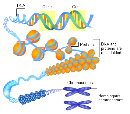 Figure 1.1 shows How a chromosome is made up of large amounts
of coiled DNA.
Figure 1.1 shows How a chromosome is made up of large amounts
of coiled DNA.
The human body is compiled of millions and millions of cells, each of these cells contain a control centre called the nucleus. This nucleus contains capillaceous structures called chromosomes, which are made up of Deoxyribonucleic Acid (DNA), see figure 1.1.[1] Each cell contains a profuse amount of DNA, so much so that if the human cell were enlarged to the size of a common tablet, our DNA would be up to 10,000 meters long![2] This DNA is wound, very tightly, around histone (proteins) in order for it to fit inside the nucleus of the cell; the structure formed after lots and lots of winding is called a 'chromosome'. Each cell contains 46 chromosomes, however there are only 23 possible chromosomes for the body to have. This is because there are two copies of each chromosome in each cell, which pair up with each other (for example, each cell contains 2 'chromosome 1's' but they form a pair, therefore there is one pair of chromosome 1's).
The only cells that do not have 23 pairs of chromosomes are the egg and sperm; they are haploid cells (only containing one set of chromosomes). This is so that when the two cells meet up and join together they make one diploid cell (containing one set of chromosomes from each parent), which is referred to as a zygote. These sex cells are synthesised in the gametes, through a process called meiosis. Meiosis is a two-step process, which reduces the number of chromosomes to 46 single, to 23 single chromosomes per cell – this forms sperm and egg cells.[3]
The only cells that do not have 23 pairs of chromosomes are the egg and sperm; they are haploid cells (only containing one set of chromosomes). This is so that when the two cells meet up and join together they make one diploid cell (containing one set of chromosomes from each parent), which is referred to as a zygote. These sex cells are synthesised in the gametes, through a process called meiosis. Meiosis is a two-step process, which reduces the number of chromosomes to 46 single, to 23 single chromosomes per cell – this forms sperm and egg cells.[3]
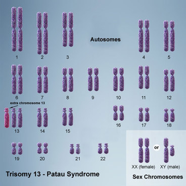 Figure 1.2 shows the third copy of the 13th Chromosome.
Patau Syndrome, or Trisomy 13, occurs when there are three copies of the 13th chromosome rather than only two copies (see figure 1.2). This occurs when on of the parents sex cells did not complete the process of meiosis properly and as a result have had 24 chromosomes in the sex cell rather than the usual, 23.[4] However having an extra copy of the 13th trisomy is not the only way for a foetus to develop patau syndrome. The characteristics categorized, as Patau syndrome, can also be a result of translocation (mispositioning). In this case, a portion of chromosome 13 is exchanged with a portion of another chromosome. Therefore a portion of each chromosome is ‘translocated’. This is the only form of Patau syndrome that can be inherited.[5]
There are three forms of patau syndrome, these include:
1. Trisomy 13 - The third chromosome 13 is present in all cells.
2. Trisomy 13 Mosaicism - The third chromosome 13 is shown in some of the cells.
3. Partial Trisomy - There is only part of a third chromosome 13, appearing in the cells.
There are three forms of patau syndrome, these include:
1. Trisomy 13 - The third chromosome 13 is present in all cells.
2. Trisomy 13 Mosaicism - The third chromosome 13 is shown in some of the cells.
3. Partial Trisomy - There is only part of a third chromosome 13, appearing in the cells.
A Little Bit Deeper...
Patau syndrome is a somewhat rare disorder, occurring in about 1 out of every 10,000 live births annually, worldwide. This rate, however, does not account for the miscarriages and stillborn babies that do not survive until full term.[5] The condition does not appear to affect a particular race or sex more prominently than another – it seems to be a sporadically spread condition. The risk of having a second child with trisomy 13 increases by 1%, sequentially with each birth. [7]
Unfortunately, it is said that roughly 50% of trisomy 13 babies will die within the first month of life and of the remaining 50%, 40% will die within the first year of life. This does mean that 5-10% will live past a year. It is impossible to determine how long for, as it becomes a case-by-case situation after the first year of life. [7] It is extremely rare for a person diagnosed with patau syndrome to make it to adulthood, however there is one known case who lived until they were 33 years of age.[5]
Survivors often have profound disabilities both mentally and physically; the severity of this varies depending on the circumstances of condition. It is hard to place a limit on the capabilities and future implementations of the condition as they are all determined by the severity of the condition but children may be able to walk without a walker. They may even be able to understand words, phrases, obey simple commands, use minimal words/signs, and recognise and interact with other people.[5]
Causes
Occasionally an error, called nondisjunction, occurs in the process of meiosis and one of the parent’s sex cells have an extra chromosome. Instead of having the required 23 chromosomes, it now has 24. This, when added to the other parents sex cells gives the zygote a total of 47 chromosomes. It should only have 46. In the condition of patau syndrome, this is what has occurred. Either the sperm or egg has contained two copies of the chromosome 13, rather than the required one copy. Either the egg or the sperm cell could contain the extra copy of chromosome 13 however it is said that roughly 1 out of every 3-4 eggs contain an extra chromosome. This number increases with age; a woman who is over the age of 35 is at a greater risk of having a child with a chromosomal aberration.[6]
The risk of having a second child with trisomy 13 increases by 1%, sequentially with each birth. [7]
The risk of having a second child with trisomy 13 increases by 1%, sequentially with each birth. [7]
Diagnosis
Patau Syndrome can be determined both in prenatal and postnatal circumstances; however no cases can actually be diagnosed until a chromosomal analysis has taken place. While it is possible to diagnose after birth, approximately 90% of cases are diagnosed prenatally.[8]
Prior to birth, Trisomy 13 is generally suspected through a detailed foetal ultrasound, and then verified by one of the following processes.
Amniocentesis - Taking cells from the amniotic fluid and testing them.
Chorionic Villus Sampling (CVS) – taking cells from the placenta and testing them.
If the syndrome is suspected after birth, Trisomy 13 can easily be detected through a physical examination however a small blood sample is also taken to confirm. A chromosome analysis, whether it is done through amniocentesis, CVS or blood sampling, has an accuracy rate of 99.9%.[6]
References
[1] Genetics Home Reference, (2014). What is a chromosome?. [online] Available at: http://ghr.nlm.nih.gov/handbook/basics/chromosome [Accessed 12 Aug. 2014].
[2] Genetic Science Learning Centre, (2014). What is a Chromosome?. [online] Virtual.unal.edu.co. Available at: http://www.virtual.unal.edu.co/cursos/ingenieria/2001832/lecturas/chromosome.swf [Accessed 12 Aug. 2014].
[3] Genetics Home Reference, (2014). How do cells divide?. [online] Available at: http://ghr.nlm.nih.gov/handbook/howgeneswork/cellsdivide [Accessed 12 Aug. 2014].
[4] Haldeman-Englert, C. (2013). Trisomy 13: MedlinePlus Medical Encyclopedia. [online] Nlm.nih.gov. Available at: http://www.nlm.nih.gov/medlineplus/ency/article/001660.htm [Accessed 12 Aug. 2014].[5] Johnson, P. (2014). Patau Syndrome - baby, symptoms, average, Definition, Description, Demographics, Causes and symptoms. [online] Healthofchildren.com. Available at: http://www.healthofchildren.com/P/Patau-Syndrome.html [Accessed 15 Aug. 2014].
[6] Barlow-Stewart, K. (2012). Trisomy 13 - Patau Syndrome. 1st ed. [ebook] Centre for Genetics Education, pp.1,2,3. Available at: http://www.genetics.edu.au/Publications-and-Resources/Genetics-Fact-Sheets/FactSheet29PatauSyndrome.pdf [Accessed 14 Aug. 2014].
[7] Chitayat, D. (2011). Trisomy 13 (Patau Syndrome). [online] Aboutkidshealth.ca. Available at:http://www.aboutkidshealth.ca/EN/HEALTHAZ/CONDITIONSANDDISEASES/GENETICDISORDERS/Pages/trisomy-13-patau-syndrome.aspx [Accessed 12 Aug. 2014].
[8] Brochures.mater.org.au, (2014). Patau Syndrome—Trisomy 13. [online] Available at: http://brochures.mater.org.au/Home/Brochures/Mater-Mothers-Hospital/Patau-Syndrome-Trisomy-13 [Accessed 14 Aug. 2014].
